Romanian Journal of Neurology Logo
  • Home
  • Aims & Scope
  • Standards
    • Editorial policies
    • Instructions for authors
    • Peer review process
    • Guidelines for Reviewers
    • Ethics and Malpractice policies
    • Official Journal’s protocols & statements
    • Open Access
    • Fees
  • EDITORIAL COUNCIL
  • Peer Review Council
  • EMC | CME
  • How to
  • SUBSCRIBE
  • Menu

Author archives

  • About
  • Latest Posts

Kishor V. Hegde

Latest posts by Kishor V. Hegde (see all)

  • CAVERNOMA OF CERVICOMEDULLARY REGION PRESENTING WITH HEMIHYPESTHESIA - 15/09/2015
  • PICTORIAL REVIEW ON CONSTELLATION OF IMAGING FINDINGS OF BRAIN DEATH ON 3T MRI - 14/09/2015

Articles signed on Romanian Journal of NEUROLOGY:

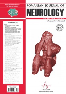
CAVERNOMA OF CERVICOMEDULLARY REGION PRESENTING WITH HEMIHYPESTHESIA

SELECT ISSUE

Romanian Journal of Neurology, Volume XIII, No. 4, 2014
ISSN 1843-8148  |  e-ISSN 2069-6094
ISSN-L 1843-8148
DOI: 10.37897/RJN

Indexed

DOI - Crossref
Similarity Check by iThenticate, worldwide No 1 professional plagiarism checking system
DOAJ
Scopus
NLM Catalog
Ebsco Host - Medline
Google Academic
Semantic Scholar

HIGHLIGHTS

ICMJE- Recommendations

Read the Recommendations for the Conduct, Reporting, Editing, and Publication of Scholarly work in Medical Journals.

Promoting Global Health

The published medical research literature is a global public good. Medical journal editors have a social responsibility to promote global health by publishing, whenever possible, research that furthers health worldwide.

CAVERNOMA OF CERVICOMEDULLARY REGION PRESENTING WITH HEMIHYPESTHESIA

Umamaheswara V. Reddy, Amit Agrawal, N.S. Sampath Kumar, Gauri Rani Karur and Kishor V. Hegde

ABSTRACT

Medullary cavernomas only contribute to 5% of brain stem cavernomas, various clinical presentations for medullary cavernomas have been described like intractable hiccups, dysphagia, hemiparesis, anorexia nervosa and hemihypesthesia. Exact pathomechanism for developing sensory symptoms has not been described earlier. Pressure effects due to changing dynamics in this cavernous malformation resulting in mass effect over the traversing sensory tract fibers is the possible mechanism. Differentials of cavernoma should be considered for any case presenting with hemihypesthesia. Surgery should be done only when there is appropriate indication, as surgery itself causes significant morbidity.

Keywords: cavernoma, cavernous angioma, medulla oblongata, hemihypesthesia

Full text | PDF

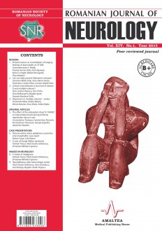
PICTORIAL REVIEW ON CONSTELLATION OF IMAGING FINDINGS OF BRAIN DEATH ON 3T MRI

SELECT ISSUE

Romanian Journal of Neurology, Volume XIV, No. 1, 2015
ISSN 1843-8148  |  e-ISSN 2069-6094
ISSN-L 1843-8148
DOI: 10.37897/RJN

Indexed

DOI - Crossref
Similarity Check by iThenticate, worldwide No 1 professional plagiarism checking system
DOAJ
Scopus
NLM Catalog
Ebsco Host - Medline
Google Academic
Semantic Scholar

HIGHLIGHTS

ICMJE- Recommendations

Read the Recommendations for the Conduct, Reporting, Editing, and Publication of Scholarly work in Medical Journals.

Promoting Global Health

The published medical research literature is a global public good. Medical journal editors have a social responsibility to promote global health by publishing, whenever possible, research that furthers health worldwide.

PICTORIAL REVIEW ON CONSTELLATION OF IMAGING FINDINGS OF BRAIN DEATH ON 3T MRI

Umamaheswara V. Reddy, Praveen Kumar Kolla, Amit Agrawal, Kishor V. Hegde, Rakesh Borrugada and Priya Malpani

ABSTRACT

Brain death is physiological loss of function of all intracranial structures above foramen magnum. Imaging is ancillary and are not substitute for clinical diagnosis of brain death. MRI can be useful supplementary diagnostic test for diagnosing brain death especially in cases where bed side clinical tests cannot be performed. No single sign in MRI is specific for brain death. Tonsillar herniation, loss of flow void in bilateral internal carotid arteries and vertebral arteries, swollen cortical gyri, edematous, cerebellar foliae with white matter hyperintensities, effacement of the ventricular system, uncal herniation compressing over the midbrain, brain stem swelling, dilated veins showing increased susceptibility phenomenon due to increase deoxyhemoglobin, diffuse restriction in cerebral cortex are some of the feature of brain death we present the imaging signs brain which we came across in two patients.

Keywords: brain death, tonsillar herniation, brain stem reflexes, Imaging signs, 3Tesla MRI

Full text | PDF


SEARCH

STANDARDS

  • Instructions for AUTHORS
  • Peer review process

Submit article

Submit your article to the journal by using the form here:

Submit

Subscriptions

To receive the Ro Journal of NEUROLOGY click here:

SUBSCRIBE
Publicare
Abonare

plic-maileditor@rjn.com.ro

Subscribe to the Newsletter

Articles from the journal are licensed under a 
Creative Commons Attribution 4.0 International License

 Terms & Conditions

Open Access Statement

Publisher : AMALTEA Medical Publishing House

The intended audience of the site content is professionals from the medical and pharmaceutical community.
This site does not collect any personal data nor does it use cookies that might obtain such data from your browser. [ info ]
Agree
Contact

Submit ARTICLE
E-mail: editor@rjn.com.ro  |  Mobile: +4 0742.155.512, M-F 09.00-18.00 EET

SUBSCRIPTIONS
E-mail: info@amaltea.ro |  Mobile: +4 0742.155.511, M-F 09.00-18.00 EET